


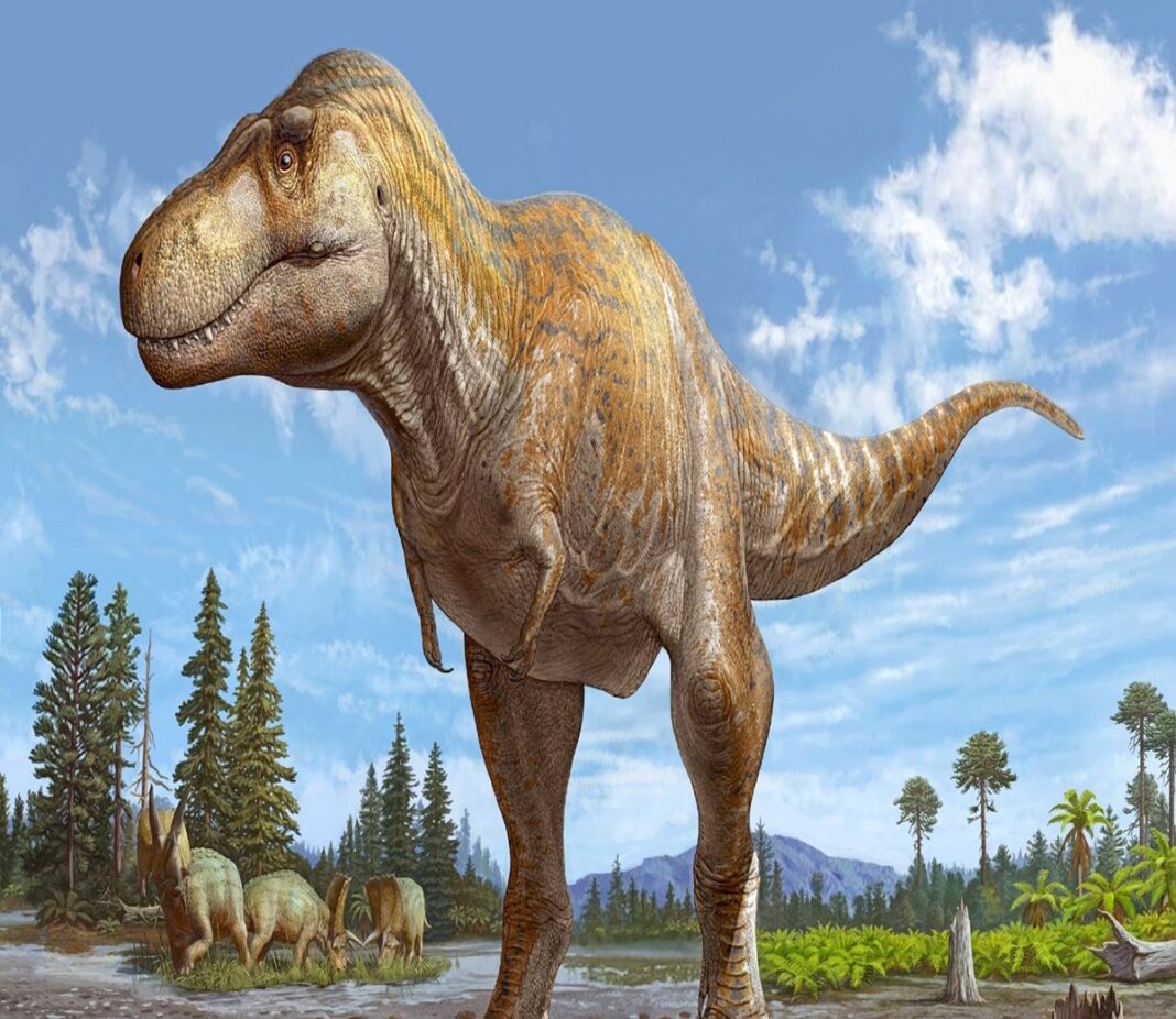
Advanced scientific imaging revealed dramatic new details regarding the death of Scotty, the largest Tyrannosaurus rex (T Rex) ever discovered. Discovered in Saskatchewan, Canada, the 66-million-year-old apex predator measured 43 feet long and weighed nearly 19,500 pounds. Non-destructive neutron analysis conducted at Oak Ridge National Laboratory uncovered a severe rib fracture sustained during a dinosaur battle. Researchers confirmed that Scotty succumbed to these combat injuries before the rib mended, preserving rare fossilized vascular tissue.
Researchers from Canada’s University of Regina and the Oak Ridge National Laboratory discovered that the 19,500-pound dinosaur suffered a severe rib fracture during a fight with another large dinosaur. High-resolution imaging confirmed that while Scotty’s body attempted to heal the broken bone by forming new blood vessels around the fracture, the injury proved fatal before full anatomical recovery occurred.
How Did T Rex Neutron Imaging Preserve Fossil?
Scientists used non-destructive neutron beams to scan the T Rex fossilized rib bone at specialized accelerator facilities. Unlike X-rays, neutrons can penetrate heavy minerals and detect lighter organic elements. The technique mapped three-dimensional networks of mineralized blood vessels and preserved soft tissue inside the ancient T. Rex bone without carving or damaging the specimen.
Scotty died in an oxygen-deprived, salty wetland environment shortly after sustaining the fatal fight injury. Waterlogged wetland soil severely inhibited microbial decomposition, allowing delicate vascular networks saturated with iron-rich blood to fossilize alongside the surrounding bone structure.
Combining particle physics with paleontology opens non-destructive avenues for examining prehistoric life. Non-invasive imaging technologies continue transforming our understanding of ancient animal biology, paleopathology, and fossil preservation mechanisms
