


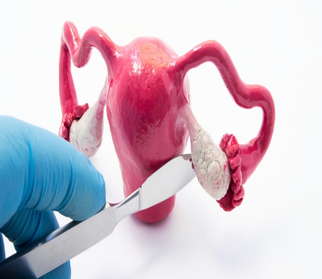
Uterus-related findings surprised medical specialists in Delhi after they identified an extremely rare anatomical anomaly during a routine fertility consultation. Clinical reports confirm that a 26-year-old man seeking treatment for primary infertility and zero sperm count was diagnosed with Persistent Müllerian Duct Syndrome (PMDS), a rare condition involving retained female reproductive structures.
Diagnostic imaging through pelvic MRI scans revealed the presence of a fully formed uterus, fallopian tubes, and associated female reproductive tissues inside the male patient’s abdominal cavity. Despite having fully virilized male external genitalia, both testes failed to descend into the scrotum, remaining trapped internally near the uterine structures. Genetic analysis confirmed a standard male 46,XY karyotype, prompting physicians to diagnose Persistent Mullerian Duct Syndrome (PMDS).
Why Does PMDS Cause A Persistent Uterus?
During typical male fetal development, the testes produce anti-Müllerian hormone, which causes early uterus and fallopian tube structures to regress. In Persistent Mullerian Duct Syndrome, the hormone may be absent or its receptors may not function properly, allowing Mullerian structures, including a uterus, to persist alongside male reproductive development. Fewer than 300 cases have been documented worldwide.
Surgeons performed a successful minimally invasive laparoscopic operation to remove the internal Mullerian organs and the abdominal testes. Histopathology tests on the excised tissue revealed early pre-cancerous germ cell changes in the undescended testicular tissue, proving that timely surgical intervention prevented malignant tumor growth.
Advanced diagnostic imaging enables early detection of rare developmental disorders. Timely surgical care protects long-term health and prevents severe cellular complications.
